Over the past thirty years, advanced medical imaging has revolutionized the field of healthcare. Previously, doctors had limited insight into the human body. However, with the advent of diagnostic imaging, medical professionals can now delve deeper into health concerns and determine the best course of action for patients using high-resolution images of soft and hard tissues. Medical facilities in the USA utilize various devices and techniques for modern medical imaging, including nuclear medicine, CT, PET, and MRI scans, ensuring accurate diagnostics through their medical imaging center.
Many physicians frequently refer their patients to a medical diagnostic imaging clinic in Suffolk County, NY, to obtain a more comprehensive understanding of health risks and disorders. Continue reading to learn more about the many imaging modalities available to medical professionals and their applications in contemporary medicine.
Ultrasound
X-rays do not come from the idea or principle of ultrasound. When someone started researching bat echolocation in the late eighteenth century, they initially recorded it. That's how ultrasound works. High-frequency sound waves are used in ultrasound to create images of soft tissues such as muscles, joints, and organs by reflecting off of tissue. It is comparable to illuminating light within the body, but it passes through the skin layers and is only visible with electronic sensors. As we know, ultrasound existed in the 20th century.
Originally used to identify brain cancers, ultrasound later emerged as the primary diagnostic modality for tracking the growth of the fetus in expectant mothers. Ultrasound has developed into a vital diagnostic tool that physicians use to assess patients and identify a range of ailments. They still use it to detect cancers but also to treat strained muscles and persistent discomfort and encourage healing. Numerous ultrasound devices, such as the GE Logiq E9, are now on the market, and they get better every year.
Radiography
X-rays are sometimes known by the prettier, more technical term, radiography. Gamma rays or electromagnetic radiation are used in radiography to provide images of the inside organs. X-rays are the most popular and widely used type of radiography. The majority of us have undergone an X-ray at some point. With this incredibly flexible imaging technique, any body area may be imaged. X-ray equipment projects high-energy waves into the body during this treatment.
Hard tissues, like bones, absorb the waves, while soft tissues, like skin and organs, do not. The X-ray machine projects the images onto a film, which darkens the unabsorbed materials and highlights the body areas that absorb the waves in white. The clarity and quality of the images have improved over time, increasing their usefulness in the diagnosis of illness.
Imaging with Magnetic Resonance (MRI)
The MRI is a common tool for all sports fans. Athletes receive them daily to determine which muscle they tore and which ligament they pulled during the game. Radio waves and magnetic fields are used in magnetic resonance imaging to examine the body's tissues, including the organs. Like other imaging modalities, MRI is painless and noninvasive. An MRI scanner, a big tube with a huge circular magnet, is needed for the treatment. While the magnet circles around the patient, capturing photographs, they lie on the table and are fed into the machine, which has a futuristic appearance. The hydrogen atoms in the body's protons align thanks to the magnet's powerful magnetic field.
The protons then rotate as a result of being exposed to radio waves. The protons relax and realign themselves when the radio waves are turned off, releasing radio waves throughout the recovery process that the system can pick up on to produce an image. The image gets crisper the faster the protons realign. When repeated imaging is required, particularly in the brain, magnetic resonance imaging (MRI) is preferred since it produces images without radiation.
Use Of Nuclear Medicine
Any use of radioactive elements in medicine is referred to as "nuclear medicine," a fairly broad word. However, imaging usually involves using radioactive tracers, substances injected or consumed to pass through the circulatory or digestive systems. An image of such systems can then be created by detecting the substance's radiation. Gamma rays are the energy trail the tracer leaves behind as it moves across the examination area. The rays are detected by a specialized camera, which then relays the data to a computer to generate an image.
Different imaging techniques cannot yield the unique information that nuclear imaging gives. It can detect fatal illnesses in the earliest stages of development. It is most frequently utilized when a medical professional wishes to view the anatomy and physiology of a bone, organ, tissue, or bodily system. The needle injection of the tracer is the sole painful part of the procedure. The usage of the tracer has no recognized negative consequences.
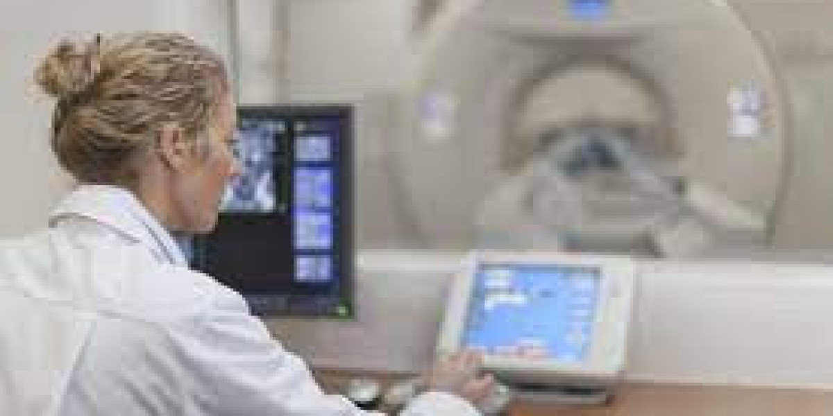
CT Scan: A Computer Tomography
Computer tomography comes last in our description of imaging technology's various kinds and uses. Since computer tomography (CT) scans also employ X-ray technology to produce cross-sectional images of the body, they are classified as radiography. They give medical professionals a clear view of organs and structures that more conventional X-ray technology would otherwise obscure. The patient lies on a motorized table inside a sizable circular chamber that resembles an MRI machine during a CT scan.
To produce a series of three-dimensional slides showing the inside of the body, the low-dose X-ray source, along with its detector or receiver, rotates around the patient. These slices can be seen individually or as a virtual, three-dimensional depiction of the impacted region when stacked. Individual slides are accessible, and the complete human body can be viewed from head to toe after they have been assembled. A physician can study a particular slide from the thousands that are cataloged to diagnose a patient.
Wrapping-Up
Medical imaging technologies have transformed the practice of medicine, enabling clinicians to diagnose diseases, plan treatments, and monitor patient progress with unprecedented precision. From the simplicity of X-rays to the sophistication of MRI and PET scans, each imaging modality offers unique advantages in different clinical scenarios. By understanding the principles and applications of these technologies, healthcare professionals can optimize patient care and improve outcomes while ensuring accessibility through affordable imaging services