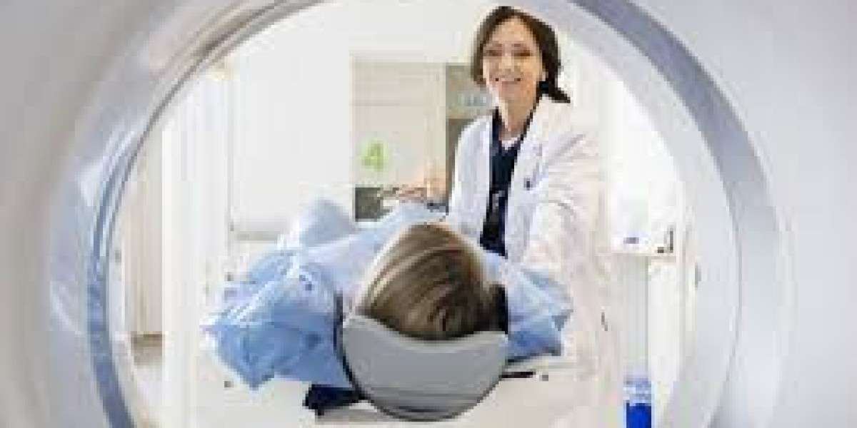
Ultrasound is a widely utilized imaging technique that employs sound waves to produce visual representations of internal body structures. Unlike other imaging methods that utilize radiation, such as X-rays or CT scans, ultrasound is a non-invasive and safe option, making it particularly valuable in various medical settings. This imaging modality has revolutionized diagnostic medicine by offering real-time visualization of organs and tissues. Its applications span multiple specialties, from obstetrics to cardiology, allowing for early diagnosis and timely intervention. This article explores the various conditions that can be diagnosed using ultrasound and highlights its significance in modern healthcare.
Ultrasound in Obstetrics and Gynecology
Pregnancy Monitoring and Fetal Health
In obstetrics, ultrasound plays a critical role in monitoring pregnancy and assessing fetal health. Routine ultrasounds are typically performed at various stages during pregnancy to evaluate fetal growth, development, and overall well-being. These scans help identify congenital anomalies, allowing for early intervention if necessary. Ultrasound can also assess placental health and measure amniotic fluid levels, ensuring that the fetus is receiving adequate nourishment and oxygen. This non-invasive imaging technique provides crucial information that helps healthcare providers make informed decisions regarding prenatal care and potential interventions.
Gynecological Conditions
Ultrasound is also instrumental in diagnosing various gynecological conditions. It can detect ovarian cysts, fibroids, and other abnormalities in the uterus. By visualizing the reproductive organs, healthcare providers can identify conditions such as endometriosis and polycystic ovary syndrome (PCOS). These conditions can significantly impact a woman’s health, causing symptoms like pelvic pain and irregular menstrual cycles. Early diagnosis through ultrasound enables timely treatment options, improving the quality of life for many women. This imaging modality serves as an essential tool in understanding and managing women's health issues.
Ultrasound in Cardiology (Echocardiography)
Heart Function and Structure Assessment
In cardiology, ultrasound, commonly known as echocardiography, is vital for assessing heart function and structure. This imaging technique helps diagnose a range of cardiovascular conditions, including heart valve issues and congenital heart defects. By utilizing ultrasound, healthcare providers can evaluate the heart's pumping ability and the condition of its valves. This information is crucial in determining appropriate treatment strategies for patients suffering from heart disease. Echocardiography provides a comprehensive overview of cardiac health, allowing for proactive management of cardiovascular conditions.
Blood Flow and Vascular Health
Ultrasound is also employed to assess blood flow in the cardiovascular system. Doppler ultrasound techniques enable healthcare providers to evaluate blood flow in arteries and veins, identifying potential issues like blockages or aneurysms. This type of imaging is essential for diagnosing conditions such as deep vein thrombosis (DVT) and peripheral artery disease (PAD). By visualizing blood flow, ultrasound can guide treatment decisions and interventions to prevent serious complications. The ability to monitor vascular health through ultrasound enhances the overall management of cardiovascular diseases.
Abdominal and Pelvic Ultrasound
Diagnosing Liver, Gallbladder, and Kidney Conditions
Abdominal ultrasound is an effective tool for diagnosing conditions affecting the liver, gallbladder, and kidneys. It can identify gallstones, liver tumors, and kidney stones, providing valuable insights into the patient's abdominal health. By visualizing these organs, healthcare providers can diagnose conditions like cirrhosis, fatty liver disease, and other abnormalities that may compromise organ function. The non-invasive nature of abdominal ultrasound makes it a preferred option for evaluating patients with abdominal pain or discomfort. This imaging modality is essential in managing gastrointestinal and urological health.
Assessing the Pancreas, Spleen, and Bladder
Ultrasound is also useful in assessing the pancreas, spleen, and bladder. It helps diagnose conditions like pancreatitis, splenic abnormalities, and bladder tumors. By examining these organs, healthcare providers can detect issues such as inflammation, infections, and masses that require further evaluation. Additionally, ultrasound plays a role in detecting urinary tract infections (UTIs) and bladder stones, aiding in timely diagnosis and treatment. This imaging technique enhances the understanding of abdominal pathology and facilitates effective management of related conditions.
Musculoskeletal Ultrasound
Diagnosing Joint and Tendon Injuries
Musculoskeletal ultrasound is increasingly used in diagnosing joint and tendon injuries. This imaging technique enables healthcare providers to visualize ligaments, tendons, and muscles, making it easier to identify conditions such as ligament tears, tendonitis, and joint inflammation. Athletes and individuals with active lifestyles benefit from ultrasound as it allows for real-time assessment of injuries. By accurately diagnosing musculoskeletal conditions, ultrasound aids in formulating appropriate treatment plans and rehabilitation strategies, helping patients return to their normal activities more quickly.
Soft Tissue and Nerve Conditions
In addition to joint injuries, ultrasound is effective in diagnosing soft tissue masses and nerve entrapment syndromes. This imaging modality can pinpoint the source of pain or dysfunction in soft tissues, such as muscles and tendons. Furthermore, it assists in diagnosing conditions like carpal tunnel syndrome, where the median nerve becomes compressed. By visualizing soft tissue structures, ultrasound provides essential information for guiding surgical interventions or conservative treatments. This versatility enhances the overall management of musculoskeletal and neurological conditions.
Breast Ultrasound
Screening for Breast Cancer
Breast ultrasound is a valuable tool in the early detection of breast cancer. It is often used as a complementary imaging modality alongside mammography. Ultrasound can help characterize breast masses and differentiate between cystic and solid lesions. By visualizing breast tissue, healthcare providers can identify abnormalities that may require further evaluation or biopsy. The use of ultrasound in breast cancer screening increases the accuracy of diagnosis, particularly in women with dense breast tissue, where mammograms may be less effective.
Differentiating Between Solid and Fluid-Filled Masses
Ultrasound is instrumental in differentiating between solid and fluid-filled masses in the breast. This distinction is critical for determining the nature of a lump and the subsequent management approach. By assessing the characteristics of breast lesions, healthcare providers can make informed decisions about the need for additional testing or treatment. Breast ultrasound enhances the diagnostic process, providing valuable insights into breast health and facilitating timely interventions when necessary.
Thyroid Ultrasound
Detecting Thyroid Nodules and Tumors
Thyroid ultrasound is essential for detecting nodules and tumors in the thyroid gland. This imaging technique allows healthcare providers to visualize the thyroid's structure and identify any abnormalities. By assessing the size and characteristics of thyroid nodules, ultrasound helps differentiate between benign and malignant growths. Early detection of thyroid tumors is crucial for effective treatment, and ultrasound plays a pivotal role in the evaluation of thyroid health. This imaging modality enhances the understanding of thyroid disorders and guides management decisions.
Role in Diagnosing Hyperthyroidism and Hypothyroidism
In addition to detecting nodules, ultrasound aids in diagnosing hyperthyroidism and hypothyroidism. By evaluating the overall health and structure of the thyroid gland, healthcare providers can identify conditions that may affect hormone production. Ultrasound can also help assess changes in the thyroid related to autoimmune conditions such as Graves' disease or Hashimoto's thyroiditis. Understanding these conditions is essential for developing appropriate treatment plans, and ultrasound serves as a vital tool in the diagnosis of thyroid disorders.
Vascular Ultrasound
Detecting Deep Vein Thrombosis (DVT)
Vascular ultrasound is particularly effective in detecting deep vein thrombosis (DVT). By using Doppler ultrasound, healthcare providers can visualize blood flow in the veins and identify the presence of blood clots. Early detection of DVT is critical for preventing serious complications such as pulmonary embolism. Ultrasound serves as a non-invasive and efficient method for evaluating venous health, providing timely interventions for patients at risk of clot formation. This imaging modality is integral to managing vascular conditions.
Assessing Carotid Artery Health
Ultrasound is also valuable in assessing carotid artery health. It helps diagnose carotid artery disease, which can lead to stroke. By evaluating blood flow and detecting blockages, vascular ultrasound plays a crucial role in determining a patient's risk for cerebrovascular events. This non-invasive imaging technique allows for early intervention and management strategies to reduce stroke risk. The ability to monitor carotid artery health enhances overall cardiovascular care.
Ultrasound in Emergency Medicine
Point-of-Care Ultrasound (POCUS) for Trauma
In emergency medicine, ultrasound is an invaluable tool, particularly through point-of-care ultrasound (POCUS). This immediate imaging technique allows healthcare providers to assess internal injuries in trauma patients. POCUS is instrumental in detecting internal bleeding, organ damage, and fractures, facilitating rapid decision-making in critical situations. By providing real-time information, ultrasound enhances patient management in emergency settings, improving outcomes for individuals with acute injuries.
Role in Rapid Diagnosis of Acute Conditions
Ultrasound also plays a significant role in the rapid diagnosis of acute conditions, such as appendicitis or ectopic pregnancy. By quickly visualizing the abdominal organs, healthcare providers can identify conditions that require urgent intervention. The ability to perform ultrasound in emergency situations allows for prompt treatment and minimizes complications. This imaging modality is crucial in emergency care, where timely diagnosis can significantly impact patient outcomes.
Limitations of Ultrasound in Diagnosis
Conditions That May Be Difficult to Detect
Despite its numerous benefits, ultrasound has limitations in diagnosing certain conditions. For instance, it may struggle to visualize structures like the lungs or bones effectively. Certain abdominal conditions may also be challenging to assess through ultrasound alone. While it provides valuable information, healthcare providers often use ultrasound in conjunction with other imaging techniques, such as MRI or CT scans, to obtain a comprehensive view of a patient's condition. Understanding these limitations is essential for interpreting ultrasound results accurately.
False Positives and Need for Further Testing
Another limitation of ultrasound is the potential for false positives, which can lead to unnecessary anxiety or additional testing. Some benign conditions may mimic more serious issues on an ultrasound image, prompting further investigations. Additionally, small tumors or abnormalities may be missed, necessitating follow-up imaging or biopsies. Awareness of these limitations helps healthcare providers interpret ultrasound results cautiously and consider the need for additional diagnostic evaluations to confirm findings.
Conclusion
In conclusion, ultrasound is a versatile and non-invasive imaging modality that plays a crucial role in diagnosing a wide range of conditions across various medical fields. From obstetrics to cardiology and emergency medicine, ultrasound provides valuable insights into patients’ health, enabling early detection and intervention. Its applications extend beyond diagnosis, guiding treatment decisions and improving patient outcomes. As technology advances, ultrasound continues to evolve, enhancing its capabilities in medical diagnostics.
Importance of Early Detection and Treatment
The significance of early detection through ultrasound cannot be overstated. Timely diagnosis of medical conditions can lead to improved treatment outcomes, reduce complications, and enhance quality of life. Regular monitoring and timely imaging evaluations allow healthcare providers to address potential health issues proactively. Patients are encouraged to undergo appropriate imaging as recommended by their healthcare providers to ensure optimal health management.
Encouragement for Patients to Consult with Healthcare Providers
Ultimately, patients should consult with their healthcare providers regarding the need for ultrasound and other imaging studies. Engaging in open communication about health concerns and the potential need for diagnostic imaging is essential for effective healthcare management. By understanding the role of ultrasound in diagnosing various conditions, patients can make informed decisions about their health and well-being, leading to better outcomes in their healthcare journey.
FAQs
What is an ultrasound, and how does it work?
An ultrasound is a non-invasive imaging technique that uses high-frequency sound waves to create visual representations of internal body structures. The process involves placing a gel on the skin and using a transducer to send and receive sound waves. These waves bounce off tissues and organs, producing images that healthcare providers can use for diagnosis.
What conditions can be diagnosed using ultrasound in obstetrics?
In obstetrics, ultrasound is crucial for monitoring pregnancy and assessing fetal health. It helps detect congenital anomalies, assess fetal growth, and monitor placental health. Routine ultrasounds can also identify complications like ectopic pregnancies or placental abruption, ensuring timely medical intervention when necessary.
How is ultrasound used in diagnosing heart conditions?
Ultrasound, particularly echocardiography, is used to assess heart function and structure. It helps diagnose conditions like heart valve issues, congenital heart defects, and cardiomyopathy. By visualizing blood flow and heart muscle activity, ultrasound enables healthcare providers to determine appropriate treatment options for patients with cardiovascular diseases.
Are there any limitations to using ultrasound for diagnosis?
Yes, while ultrasound is a valuable diagnostic tool, it has limitations. It may not effectively visualize structures like bones or lungs and can produce false positives, leading to unnecessary additional testing. Some small tumors or abnormalities may also be missed, making it essential to use ultrasound alongside other imaging modalities when needed.
ultrasound safe for all patients?
Ultrasound is generally considered safe and non-invasive, with no known harmful effects from the sound waves used. However, certain patient populations, such as those with specific allergies to contrast agents or pregnant women, may require special considerations. It's important for patients to discuss their health history with their healthcare providers to ensure the safe use of ultrasound.


