What is High Content Screening?
High content screening (HCS) is a cellular imaging-based analytical technique used to observe, analyze and quantify cellular events inside living cells in a miniaturized, high-throughput format. By capturing digital images of cells and quantifying hundreds of cellular parameters, HCS enables researchers to study complex biological processes and cell responses to various treatments or external stimuli.
Advantages of HCS Over Conventional Screening Methods
Conventional drug and compound screening methods like ELISA or reporter gene assays only provide insights into a single endpoint or read-out per assay. In comparison, HCS enables multi-parametric profiling by capturing a comprehensive panel of features per cell which can range from DNA content and mitochondrial mass to cell morphology and immunostaining patterns. This allows researchers to gain a more holistic view of intracellular responses and detect subtle phenotypic changes with high resolution. HCS is also more efficient as it can analyze thousands of samples simultaneously in microplate format compared to traditional one-by-one methods. The miniaturized, automated workflows of High Content Screening translate to significant reduction in reagent consumption and handling time.
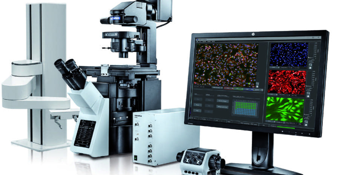
Cellular Imaging Technologies Used in HCS
Modern HCS systems utilize advanced fluorescence microscopy and image acquisition setups for rapid acquisition of high-resolution digital images. Labeling cells with fluorescent probes targeted against cellular structures or proteins of interest enables visualization of various intracellular parameters. Common labeling methods used in HCS include DAPI staining for DNA quantification, MitoTracker dyes for mitochondrial mass measurement, fluorescent conjugates against cytoskeletal or organelle proteins and immunofluorescence staining against target biomarkers. HCS systems are equipped with high throughput fluorescence microscopes, fast scanning laser confocal platforms or line-scanning abilities to rapidly capture digital images from entire microplates with high pixel density. Sophisticated image processing software then extracts quantitative readouts from each image through segmentation, object identification and measurement algorithms.
Get more insights on - High Content Screening